-
0
Patient Assessment
- 0.1 Patient Demand
- 0.2 Anatomical location
-
0.3
Patient History
- 2.1 General patient history
- 2.2 Local history
-
0.4
Risk Assessment
- 3.1 Risk Assessment Overview
- 3.2 Age
- 3.3 Patient Compliance
- 3.4 Smoking
- 3.5 Drug Abuse
- 3.6 Recreational Drug and Alcohol Abuse
- 3.7 Condition of Natural Teeth
- 3.8 Parafunctions
- 3.9 Diabetes
- 3.10 Anticoagulants
- 3.11 Osteoporosis
- 3.12 Bisphosphonates
- 3.13 MRONJ
- 3.14 Steroids
- 3.15 Radiotherapy
- 3.16 Risk factors
-
1
Diagnostics
-
2
Treatment Options
-
2.1
Treatment planning
- 0.1 Non-implant based treatment options
- 0.2 Treatment planning conventional, model based, non-guided, semi-guided
- 0.3 Digital treatment planning
- 0.4 NobelClinician and digital workflow
- 0.5 Implant position considerations overview
- 0.6 Soft tissue condition and morphology
- 0.7 Site development, soft tissue management
- 0.8 Hard tissue and bone quality
- 0.9 Site development, hard tissue management
- 0.10 Time to function
- 0.11 Submerged vs non-submerged
- 0.12 Healed or fresh extraction socket
- 0.13 Screw-retained vs. cement-retained
- 0.14 Angulated Screw Channel system (ASC)
- 2.2 Treatment options esthetic zone
- 2.3 Treatment options posterior zone
- 2.4 Comprehensive treatment concepts
-
2.1
Treatment planning
-
3
Treatment Procedures
-
3.1
Treatment procedures general considerations
- 0.1 Anesthesia
- 0.2 peri-operative care
- 0.3 Flap- or flapless
- 0.4 Non-guided protocol
- 0.5 Semi-guided protocol
- 0.6 Guided protocol overview
- 0.7 Guided protocol NobelGuide
- 0.8 Parallel implant placement considerations
- 0.9 Tapered implant placement considerations
- 0.10 3D implant position
- 0.11 Implant insertion torque
- 0.12 Intra-operative complications
- 0.13 Impression procedures, digital impressions, intraoral scanning
- 3.2 Treatment procedures esthetic zone surgical
- 3.3 Treatment procedures esthetic zone prosthetic
- 3.4 Treatment procedures posterior zone surgical
- 3.5 Treatment procedures posterior zone prosthetic
-
3.1
Treatment procedures general considerations
-
4
Aftercare
歯牙-歯肉複合体と生物学的幅径
Key points
- 生物学的幅径は、角化口腔粘膜上皮、インプラント周囲の接合上皮および上皮下結合組織により決定します。
- 歯肉とインプラント周囲粘膜では、細胞、線維、コラーゲンおよび血管の組成が異なります。
インプラント周囲粘膜と歯肉の違い
インプラント周囲粘膜は、外部の角化した口腔上皮とアバットメントに面する接合上皮により構成されます。接合上皮は軟組織の冠側縁から約2mm根尖側まで伸展していますが、インプラント周囲の歯槽頂までは、なお1.0~1.5mmの隔たりがあります。
インプラントと天然歯では生物学的幅径が異なり、インプラント周囲では>0.5mm大きく(3.8 vs 3.2mm)なります。これは、インプラント周囲粘膜では間に入る結合組織の高さが大きくなるためです。

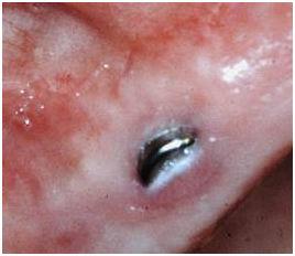
図 1: サブマージドインプラントの偶発的な粘膜穿孔。穿孔が起こると、高頻度で辺縁骨吸収が起こりますが、これは生物学的幅径を調整するためであると考えられます。
インプラント周囲の骨縁上結合組織は、天然歯と比較して線維芽細胞が少なく、コラーゲン線維を多く含みます。インプラント/アバットメント表面の付近では、辺縁骨に付着した膠原線維がインプラント/アバットメント表面と平行に走行しています。天然歯周囲の歯肉の線維は主に環状であり、歯根のセメント質ではインプラント周囲とは異なる向きで付着しています。
インプラント周囲の骨縁上結合組織を走行する血管は、天然歯周囲の同じ位置に比べると少なくなります。
インプラント周囲の結合組織は、その細胞、線維、コラーゲンおよび血管の組成から、瘢痕組織に類似しているといえます。
大規模データの解析から、インプラントの素材(チタン、ジルコニア等)や粗造度が異なっても、軽微な差こそあれ、ほぼ同じ組成パターンを示すことが分かっています。